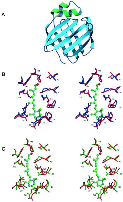
Figure 3.
(A) Ribbon drawing of the molecular model of CRBP III. Barrel forming β-strands are shown in light blue and the two α-helices are shown in green. (B) Stereoview showing the superposition of residues in proximity to the bound retinol in rat holo-CRBP I (blue colored, PDB ID code 1CRB) with the corresponding residues in human apo-CRBP III (red colored, this work). (C) Stereoview showing the superposition of residues in proximity to the bound retinol in rat holo-CRBP II (green colored, PDB ID code 1OPB) with the corresponding residues in human apo-CRBP III (red colored, this work). Vitamin A bound to CRBP I and II (ball-stick model) is shown also.

