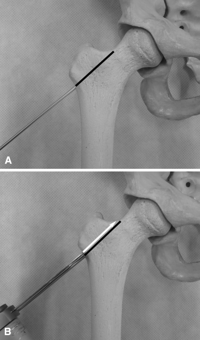
Fig. 1A–B.
(A) The guide wire is positioned at the base of the greater trochanter and then driven to the superior border of the femoral neck, as marked with the black line; and (B) the osteotomy is performed with an oscillating saw following the guide wire immediately proximal to it, as indicated by the white arrow.

