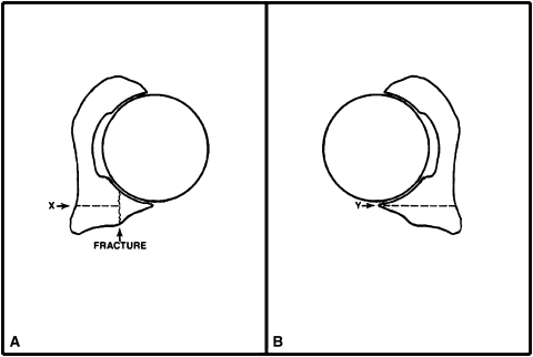
Fig. 2A–B.
The diagrams illustrate the measurement method. (A) The approximate mediolateral dimension (depth) of the smallest remaining intact posterior wall measured to the medial extent of the quadrilateral plate (X) is determined at the level of the greatest size of the posterior wall fracture fragment. (B) The percentage of fragment size is calculated from the ratio of the estimated depth of the fractured segment to the intact matched contralateral acetabular depth measured to the medial extent of the quadrilateral plate (Y) at a level comparable to that used for measurement of the fracture fragment. Y − X divided by Y multiplied by 100 provides the percentage. (Modified and reprinted with permission from Moed BR, Ajibade DA, Israel H. Computed tomography as a predictor of hip stability status in posterior wall fractures of the acetabulum. J Orthop Trauma. 2009;23:7–15.)

