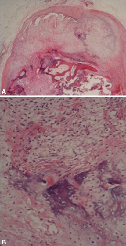
Fig. 1A–B.
(A) A histologic specimen shows a protuberance of the cellular cartilage cap with the underlying bluish stain of enchondral ossification (Stain, hematoxylin and eosin; original magnification, ×40). Deep to this, the trabecular bone (pink) is seen. (B) A histologic specimen shows the blue enchondral ossification at a higher power amid cellular cartilage and fibrous tissue (Stain, hematoxylin and eosin; original magnification, ×200).

