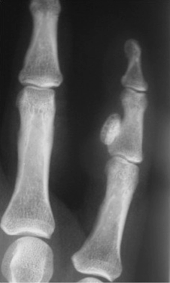
Fig. 2.

An AP radiograph shows a typical BPOP. A well-defined ossified lesion can be seen on the radiopalmer aspect of the middle phalanx of the right little finger contiguous with the cortex but without cortical/medullary infiltration or soft tissue swelling.
