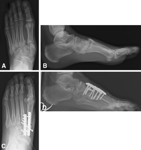
Fig. 1A−D.
(A) AP and (B) lateral radiographs are shown for a patient with a Lisfranc fracture-dislocation. There is lateral and dorsal displacement of the base of the second metatarsal with respect to the middle cuneiform. Postoperative (C) AP and (D) lateral radiographs of the same patient after ORIF are shown. A combination of dorsal plating and independent screw fixation was used to restore proper anatomic alignment to the midfoot in the coronal and sagittal planes

