Figure 2.
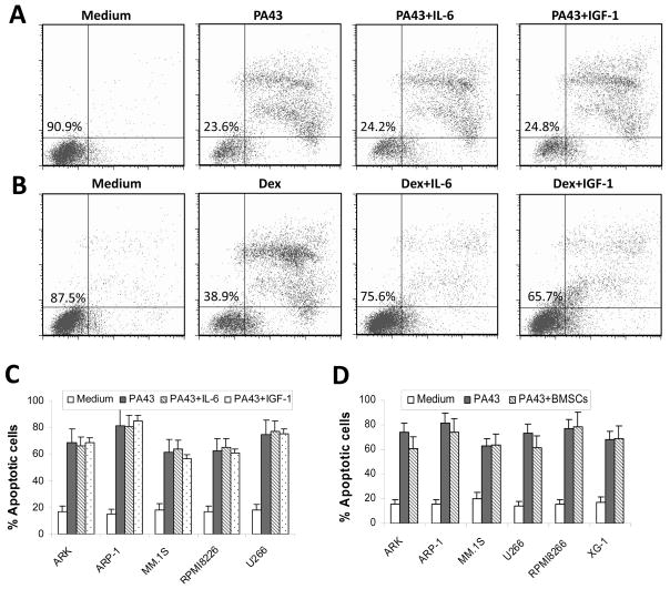
Tumouricidal activity of IgM anti-β2M mAbs is not compromised by myeloma growth factors or bone marrow stromal cells. Shown are percentages of apoptotic myeloma ARP-1 cells in 24-h cultures with (A) IgM PA43 mAb (50 μg/ml) or (B) dexamethasone (10 μM) with or without IL-6 (10 ng/ml) or IGF-I (50 ng/ml). Figures in the left quadrant represent the percentage of live cells. (C) Pooled data of apoptotic cells of five myeloma cell lines induced by IgM anti-β2M mAb with or without IL-6 or IGF-I. (D) Percentage of apoptotic cells from six myeloma cell lines in the cocultures with bone marrow stromal cells (BMSCs) with addition of IgM PA43 mAb (50 μg/ml) for 24 h. Myeloma cells cultured with medium or with the mAb without BMSCs served as controls. After coculture, myeloma cells were recovered and apoptotic cells were detected by using Annexin V-binding assay. Results of three experiments are shown.

