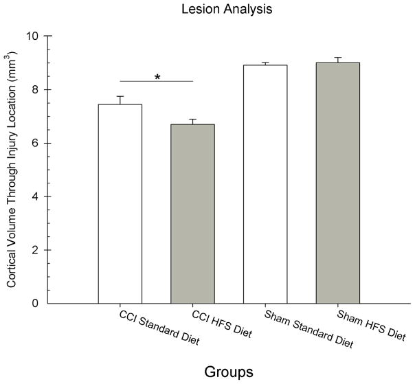
Figure 4.
The HFS diet increased the size of the injury cavity compared to the standard diet. Analysis of the remaining frontal cortical volume is shown for each group. The graph shows plotted mean (+S.E.M.) cortical volumes for each group. The HFS diet resulted in less remaining cortical tissue (i.e., larger injury cavity) compared to the standard diet (* = p < 0.01).

