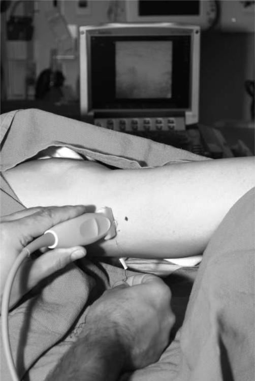
Figure 1.
Photo showing the placement of probe at the lateral aspect of the lower leg to image the peroneal nerve in cross-section at the fibular head (black dot on skin represents caudal border of the fibular head). It is important that attention be paid to ergonomics, including placement of the ultrasound screen, and that an assistant be available to help with adjustments of the ultrasound machine.

