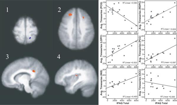
Figure 1.
Brain regions showing significant associations between self-reported physical activity and responses to heat pain in FM patients. A negative correlation was found in the post-central gyrus, extending into the superior parietal cortex (image 1). Positive correlations were found in the left and right dorsolateral prefrontal cortex (image 2), posterior cingulate cortex (image 3), and the mid to posterior insula (image 4). Images shown are with voxel-wise threshold set a p=0.005 and cluster size thresholding at 200 mm3. Functional timeseries data (average cluster values) for each individual were extracted and are shown plotted against physical activity values with the corresponding r-squared values.

