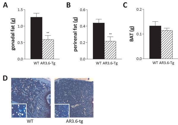
Figure 4. Decreased visceral adipose tissue mass and reduced marrow adipogenesis in AR transgenic male mice.
Individual fat depots were dissected from 6-month-old AR3.6-tg and wild-type male mice and wet tissue weights were determined. A. Gonadal (epididymal) fat, B. Perirenal fat, and C. Interscapular BAT. Data presented as mean ± SEM; n = 15-25, **, p < 0.01. D. Histological analysis after toluidine blue staining of the bone marrow compartment from the metaphyseal region of the femur in wild-type and AR3.6-tg males. Adipocytes are represented by the large empty spaces in the marrow after processing and staining. Total magnification at 50 X; inset image at 100 X total magnification.

