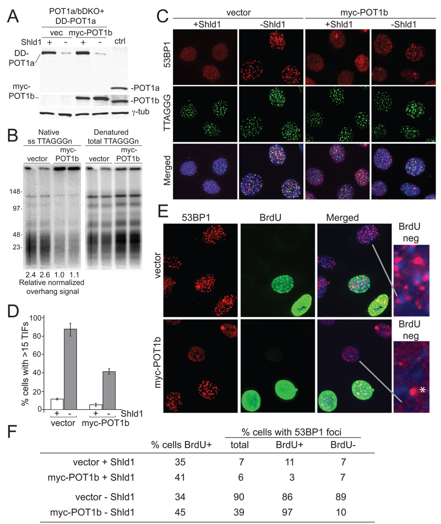
Figure 6. Different requirements for POT1-mediated telomere protection in G1 and S/G2.
(A) Immunoblotting comparing the expression of DD-POT1a and myc-POT1b in clone c223 to the endogenous POT1a and POT1b in the parental POT1a/bDKO cells (lane marked ctrl). Myc-POT1b (or empty vector) transduced c223 cells were incubated in the presence or absence of Shld1 for 16 hours and processed for immunoblotting. POT1a was detected with Ab 1221p; POT1b was detected with Ab 1223. (B) Rapid restoration of telomeric overhangs by myc-POT1b. Cells were collected at 48 hrs after infection with myc-POT1b (or the empty vector) and processed for telomeric DNA analysis in duplicate. Overhang signals were quantified with ImageQuant software and normalized to the denatured TTAGGG signal in the same lane. The numbers below the lanes show relative values of the normalized overhang signals with the value for lane 3 set to 1.0. (C) Effect of myc-POT1b expression on the TIF response after DD-POT1a depletion. Cells were collected as described in panel A and processed for IF-FISH as in Figure 1. (D) Quantification of the TIF response in C. Bars show average values and standard deviations derived from three experiments (>100 nuclei/experiment). (E) 53BP1 and BrdU co-staining in c223 cells with and without myc-POT1b. Asynchronous cultures were incubated without Shld1 for 4 hrs in the presence of 10 µM BrdU and then processed for IF for 53BP1 (red) and BrdU (green). Enlarged images show examples of 53BP1 pattern in nuclei lacking BrdU. Asterisk indicates the type of single 53BP1 foci often observed in untreated G1 cells. The foci are not indicative of a telomeric DNA damage response. (F) Quantification of the 53BP1 foci in BrdU positive and negative c223 cells with or without myc-POT1b. Cells were processed as in panel E and examined for 53BP1 foci. Cells with 15 or more 53BP1 foci were scored positive and evaluated for BrdU staining. Values are based on 150–250 cells. Similar data was obtained in a second independent experiment.

