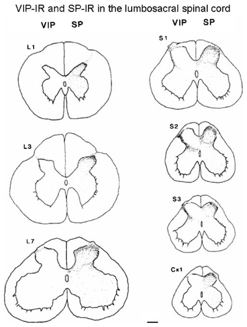
Fig. 2.
Camera lucida drawings of VIP- and substance P (SP)-IR at various levels of the lumbosacral and coccygeal spinal cord. Note that VIP-IR is present at all levels in Lissauer’s tract and lamina I of the dorsal horn, but was most prominent in the sacral segments on the lateral side of the dorsal horn. SP-IR is distributed throughout laminae I–III at all segmental levels. Calibration bar=200 µm. Reproduced with permission from Kawatani et al. (1985a)

