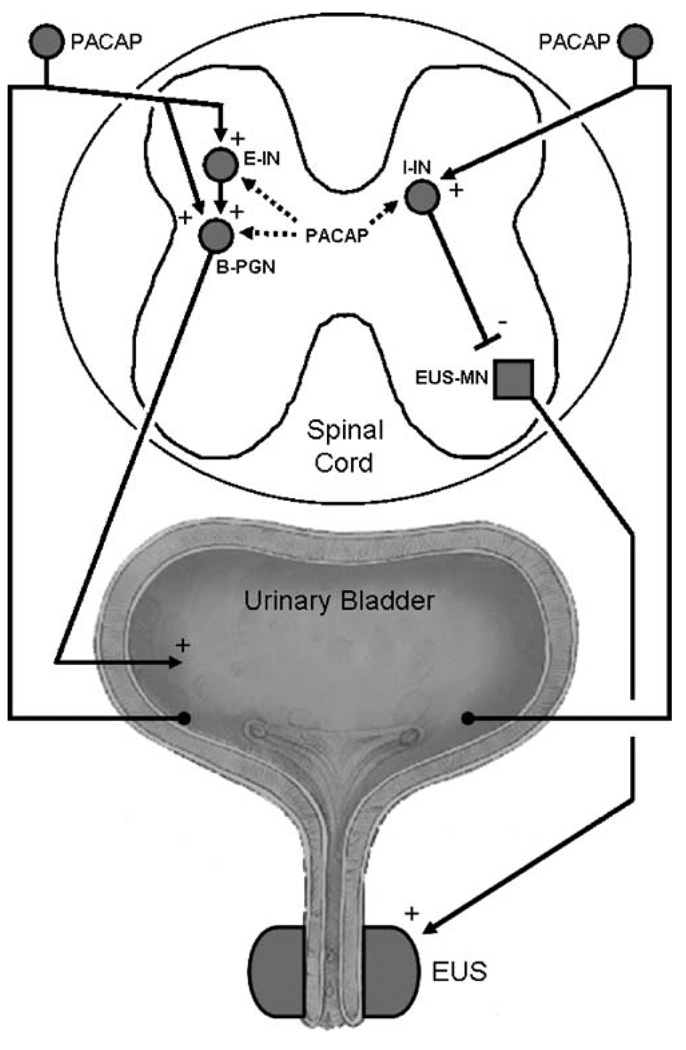
Fig. 5.
Diagram showing the possible sites in the spinal cord where PACAP acts to modulate reflex bladder and external urethral sphincter (EUS) activity in the chronic spinal cord injured rat. Left side shows the micturition reflex pathway consisting of afferent limb passing from the bladder through the dorsal roots, making excitatory synaptic connections with an excitatory interneuron (E-IN) or with a bladder preganglionic neuron (B-PGN). PACAP may function as an excitatory transmitter in bladder afferent neurons. The right side shows a putative inhibitory pathway that controls the activity of the external urethral sphincter (EUS). This pathway consists of an afferent limb arising in the bladder which synapses on an inhibitory interneuron (I-IN) which in turn makes inhibitory synaptic connections with an external sphincter motoneuron (EUS-MN) that provides the excitatory input to the EUS. The I-IN is thought to be located in the dorsal commissure region of the spinal cord. PACAP administered intrathecally could excite the bladder by activating E-INs or B-PGNs and could inhibit the EUS by activating I-INs. Excitatory pathways from the bladder to the EUS which mediate detrusor-sphincter dyssynergia and which are also prominent after spinal cord injury are not shown in the diagram. Plus and minus signs indicate excitatory and inhibitory synapses, respectively

