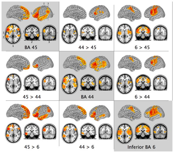
Figure 1.
Patterns of group-level resting state functional connectivity associated with the three manually-selected ventrolateral frontal ROIs, and the results of direct contrasts between them (Z > 2.3; cluster significance p < 0.05, corrected). Images are in MNI152 space and are shown according to neurological convention (right is right).

