
Figure 6.
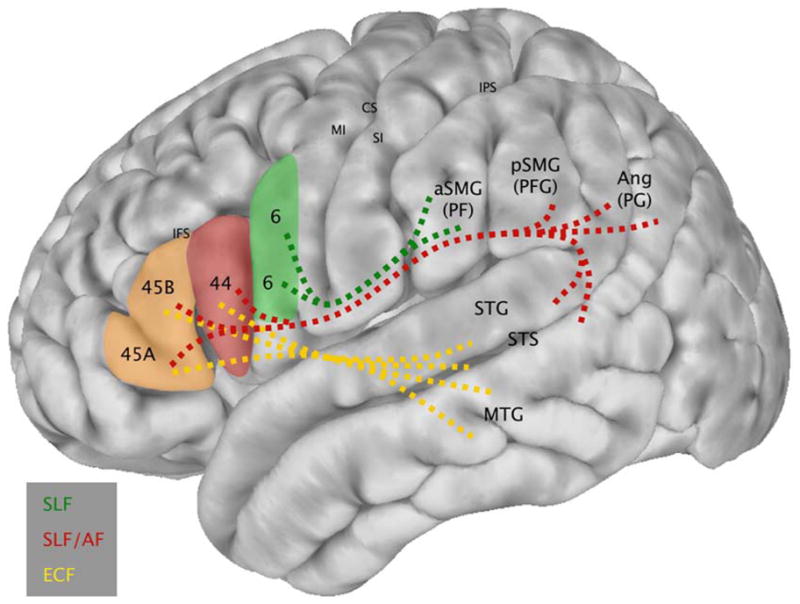
Schematic diagram integrating the observed patterns of functional connectivity between BAs 6, 44, and 45 and perisylvian parietal and temporal regions (i.e., the results of the present study) with information concerning the white matter tracts that join these regions, derived from experimental anatomical tracer studies in the macaque monkey that can demonstrate the precise origin, trajectory and termination of axonal fiber systems (Petrides & Pandya, 2009). Lines are dashed to indicate that the white matter pathways underlying the observed functional connectivity are hypothesized, but not measured directly in the present study. SLF: Superior Longitudinal Fasciculus; AF: Arcuate Fasciculus; ECF: Extreme Capsule Fasciculus.