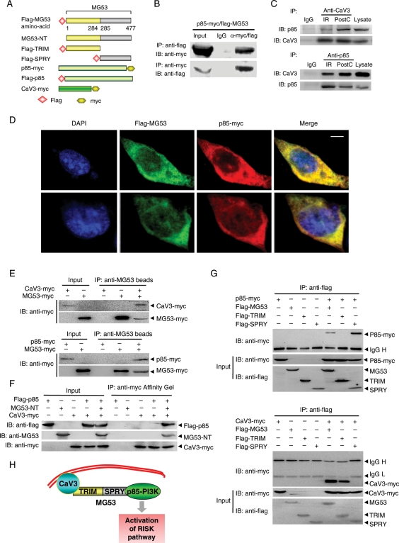
Figure 4.
MG53 interacts with p85 subunit of PI3K as well as CaV3. (A) Schematic diagram of structure of the plasmids expressing flag-MG53, MG53-NT, flag-MG53-TRIM and flag-MG53-SPRY, p85-myc, flag-p85, or CaV3-myc. (B) Co-IP of flag-MG53 and p85-myc in lysates of HEK293 cells (n = 4). (C) Co-IP of p85-PI3K and CaV3 in the lysates of the perfused mouse hearts subjected to IR with or without PostC treatment (n = 4). (D) Confocal immunofluorescence co-staining to visualize the colocalization of flag-MG53 (green) and p85-myc (red) in HEK293 cells (Scale bar denotes 5 µm). (E and F) Representative blots of pull-downed MG53-myc recombinant protein with CaV3-myc or p85-myc protein, and CaV3-myc with flag-p85 in the presence or absence of MG53-NT recombinant protein, respectively. (G) Co-IP of MG53-TRIM or MG53-SPRY with p85-PI3K-myc or CaV3-myc in lysates of HEK293 cells (n = 4). (H) Schematic presentation to show the role of MG53 in tethering CaV3 and p85-PI3K and subsequent activation of the RISK pathway.

