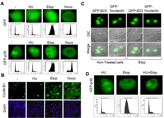
Figure 1.
Nucleolar delocalization of the HTLV-1 p30 protein. (A) Nucleolar delocalization of p30 in cells treated with etoposide. HeLa cells were transfected with GFP or GFP-p30 expression vectors, and 10 hours later were either left untreated or treated for 24 hours with hydroxyurea, etoposide, or nocodazole to synchronize them, respectively, in G1, G2, and mitosis. Cells were then fixed, mounted, and the localization of p30 was observed by epifluorescence microscopy (upper panels). Distribution of treated cells into cell-cycle phases was also accessed by flow cytometry (lower panels) or by immunofluorescence staining of cyclin B1, a cell-cycle marker exclusively expressed in G2/M (B). (C) Specificity of nucleolar delocalization of p30. HeLa cells were transfected with GFP-B23 or GFP-nucleolin expression vectors (2 structural nucleolar proteins) for 10 hours, and then either left untreated or treated with etoposide for 24 hours. The nucleolar localization of GFP-B23 and GFP-nucleolin was observed in the same conditions described in panel A. (D) Nucleolar delocalization of p30 was independent of the cell cycle and caused by etoposide treatment. HeLa cells were transfected with GFP-p30 and treated with hydroxyurea, etoposide, or hydroxyurea, followed by etoposide (top row) and cell-cycle analysis showing the distribution of cells treated under these conditions (bottom row).

