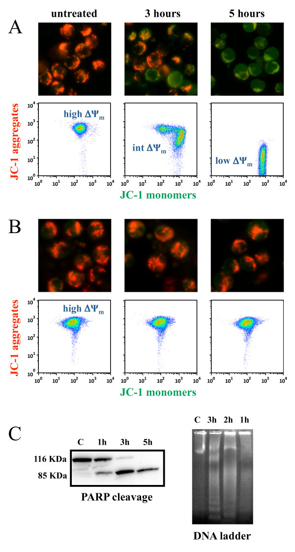
Figure 7.
α-bisabolol-induced mitochondrial damage in primary leukemic blasts. Cells were stained with JC-1. In non-damaged cells, JC-1 forms red-emitting aggregates in the mitochondrial matrix. A loss of red fluorescence and an increase in cytoplasmic green-emitting monomers signal the disruption of the mitochondrial transmembrane potential (ΔΨm). (A) The representative case Ph-B-ALL #01 is shown out of the 6 leukemias tested. Microscopy (magnification, × 400). Whereas untreated leukemic blasts showed well-polarized mitochondria marked by punctated red fluorescent staining, blasts treated with 40 μM α-bisabolol had staining that was quite completely replaced by diffuse green fluorescence, indicating loss of ΔΨm. Flow cytometry. Untreated blasts with well-polarized mitochondria localized in the upper region of the plot (high ΔΨm). Blasts exposed to 40 μM α-bisabolol shifted right and downward (intermediate and low ΔΨm), due to the progressive dislocation of JC-1 from the mitochondria to the cytoplasm, which signaled the disruption of the mitochondrial ΔΨm. (B) Both untreated and α-bisabolol-treated normal lymphocytes used as a negative control maintained well-polarized mitochondria and did not undergo apoptosis. Apoptosis of leukemic blasts was also documented by (C) PARP cleavage and DNA laddering in the same representative case depicted in (A).

