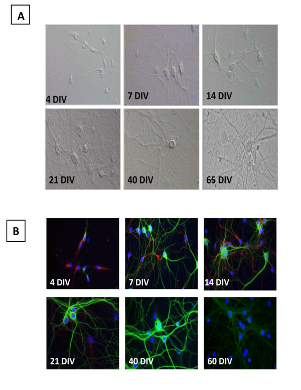
Figure 1.
Rat hippocampal neurons show distinct morphology (MAP-2; green) at different time points (A) and stop expressing the intermediate filament VI, nestin (red), over time (B). Cell bodies are stained with Hoerscht. A. Young (4 DIV) cells display small soma and short, thin neurites. As time passes the somas become larger and more defined, while neurites elongate (7 - 40 DIV). Once mature (21-40 DIV), the network takes on a progressively more intricate and bundled appearance. At 65 DIV, excessive bundling is present. B. The expression of nestin degrades over time (4 DIV- 21 DIV). MAP-2 expression appears to gain intensity up to 40 DIV. At 60 DIV, the MAP-2 expression becomes faded, and takes on a "beaded" appearance on some neurites.

