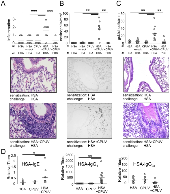
Figure 1. Parallel exposure of mice to CPUV and HSA induces allergic airway sensitization.
A: Inflammatory scores of H&E stained lung sections of mice after sensitization and challenge. Shown below are representative sections. B: Staining of lung sections for eosinophil-specific peroxidase. Total numbers of eosinophils were related to the total area of the section. Shown below are representative sections. C: Goblet cells visualized by PAS staining. Total numbers of goblet cells were related to the total length of bronchial basal membrane in the section. Representative sections are shown below. D: HSA-specific IgE, IgG1 and IgG2a titers. *p≤0.05, **p≤0.01, ***p≤0.001.

