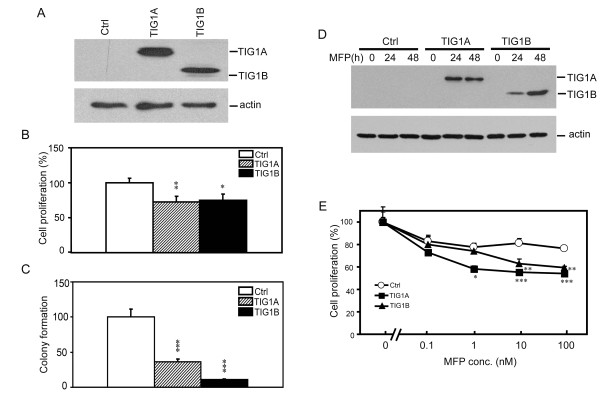
Figure 2.
Suppression of HCT116 cell growth by TIG1A and TIG1B. (A) Western blot analysis of TIG1 isoforms in transiently transfected HCT116 cells. Cells were transiently transfected with the indicated expression vectors for 24 h. Expression of TIG1A and TIG1B was detected using an anti-myc antibody. β-actin expression served as a loading control. (B) Analysis of the effects of TIG1 isoforms on cell proliferation using the WST-1 cell proliferation assay. Cells were transiently transfected with the indicated constitutive expression vectors for 48 h after which cell proliferation was measured. Data represent the mean and SD of the percentage of cell proliferation performed in triplicate and are representative of two independent experiments. (C) The effects of TIG1 isoforms on HCT116 colony formation. Cells were transiently transfected with the indicated constitutive expression vector, and colony formation was determined. Data represent the mean and SD of percentage colony formation normalised to the control performed in triplicate. Results are representative of three independent experiments. (D) Analysis of TIG1A and TIG1B expression in stable cell lines. TIG1A, TIG1B, or control stable clones were incubated in the presence or absence of 5 nM MFP for 0 - 48 h, and expression of TIG1 isoforms was analysed using Western blots analysis and anti-V5 antibody. β-actin expression served as a loading control. (E) Effects of TIG1 isoform expression on the growth of TIG1 stable cells. TIG1 and control stable clones were incubated with various concentrations of MFP or vehicle alone for 48 h. Cell proliferation was measured using the WST-1 assay. Data represent the mean and SD of percentage of cell proliferation performed in triplicate. Results are representative of three independent experiments. *, P < 0.05; **, P < 0.01; ***, P < 0.001 compared with control stable cells incubated with the indicated concentration of MFP. Ctrl, control.

