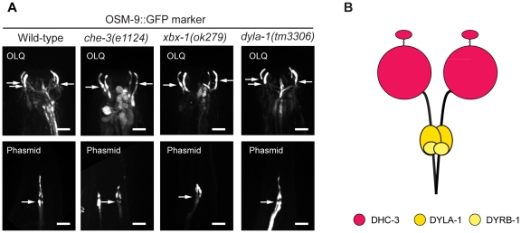
Figure 5. The gross structure of cilia associated with OLQ neurons is independent of conventional IFT-dynein.
(A) A ciliary membrane marker, OSM-9::GFP, was used to visualize the structure of cilia of OLQ neurons and of phasmid neurons. Compared to those of wild type, the ciliary structure in mutants of two essential components of conventional dynein, che-3(e1124) and xbx-1(ok279), is intact in the OLQ neurons, while truncated in the phasmid neurons. The ciliary structure is not grossly perturbed in the dyla-1(tm3306) mutant. The arrows point toward the ciliary base and the horizontal bars represent 5 µm in all panels. (B) A hypothetical new IFT-dynein could exist in C. elegans OLQ neurons.

