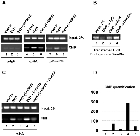
Figure 4. EVI1 and de novo DNA methyltransferases occupy the regulatory region of miR-124-3.
A. ChIP assay was performed with chromatin fragments obtained from 293T cells transiently transfected with empty vector (lanes 1, 4, and 7), EVI1 (lanes 2, 5, and 8) and EVI1-(1+6Mut) (lanes 3, 6, and 9). For the ChIP, we used IgG (lanes 1 to 3) or anti-HA (lanes 4 to 6) or anti-Dnmt3b (lanes 7 to 9) antibodies. Transfected EVI1 (lane 5, lower panel) and endogenous Dnmt3b (lane 8, lower panel) are present together on the putative miR-124-3 promoter. In contrast, when EVI1 is not expressed (lanes 4 and 7) or is mutated (lanes 6 and 9) Dnmt3b is less capable of binding to chromatin. Lanes 1–3 represent negative control with unspecific IgG. B. Dnmt3a is also enriched at the putative miR-124-3 promoter in EVI1-transfected cells (lane 4). ChIP assay was performed with chromatin fragments derived from 293T cells transiently transfected with EVI1. C. Cooperation between EVI1 and Dnmt3a in promoter occupancy. 293T cells were transiently transfected with the empty vector (lane 1), EVI1 and EVI1-(1+6Mut) alone (lanes 2 and 3) or in combination with Dnmt3a (lanes 4 and 5). The cells were used for chromatin fragments isolation/ChIP with anti-HA antibody. Normal EVI1 and Dnmt3a are more efficient in ChIP (compare lanes 2 and 4). D. ChIP quantification. The signal for cells transfected with the empty vector was arbitrarily taken as 1. Lanes numbering in C and D is the same.

