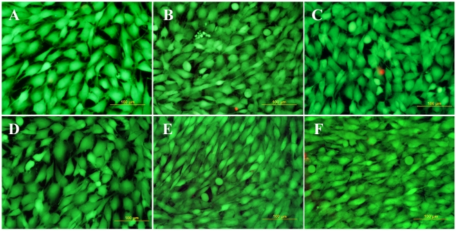
Figure 5. Fluorescence staining of MG 63 cells with a LIVE/DEAD viability/cytotoxicity kit (Invitrogen, Molecular Probes, U.S.A.) on day 7 after seeding on a microscopic glass coverslip (A) standard polystyrene cell culture dish (B), undoped NCD films (C), NCD films doped with boron in concentrations of 133 ppm (D) 1000 ppm (E) and 6700 ppm (F).
Viable cells are stained in green with calcein, dead or damaged cells in red with ethidium homodimer-1. Olympus IX 51 epifluorescence microscope, DP 70 digital camera, obj. 20×, bar = 100 µm.

