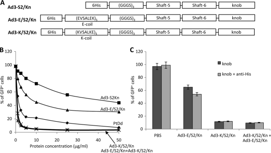
Fig. 3.
Ad3 fiber knob dimerization via E/K coils. (A) Schematic structure of recombinant Ad3 fiber knob proteins containing an N-terminal His tag, dimerization domains (E coil or K coil [17]), a flexible linker, two fiber shaft motifs (fifth and sixth), and the Ad3 fiber knob domain. Ad3-S2/Kn is a fiber that lacks the dimerization domains. (B) Competition of Ad3-GFP transduction. HeLa cells were incubated with increasing concentrations of PtDd and different Ad3 fiber knobs for 60 min and then infected with Ad3-GFP at an MOI of 100 PFU/cell for 60 min, after which the viruses were removed and new medium added. GFP fluorescence was measured 18 h later. n = 3. Shown are average values of percent GFP-positive cells. Ad3-K/S2/Kn+Ad3-E/S2/Kn is a 1:1 mixture of both fiber knobs. PtDd versus Ad3-E/S2/Kn, P = 0.074; PtDd versus Ad3-K/S2/Kn + Ad3-E/S2/Kn, P = 0.03; Ad3-K/S2/Kn versus Ad3-K/S2/Kn + Ad3-E/S2/Kn, P = 0.62. (C) Cross-linking of Ad3 fiber knobs with anti-His antibodies. Five μg/ml of Ad3 fiber knobs was incubated with 20 μg/ml of mouse anti-His MAb at room temperature for 60 min and then added to 1 ×10⁵ HeLa cells. After 60 min of incubation, 100 PFU/cell of Ad3-GFP virus was added and GFP was analyzed as described for panel B. The difference between knob and knob plus anti-His is significant for Ad3-E/S2/Kn (P < 0.05) but not for the other samples.

