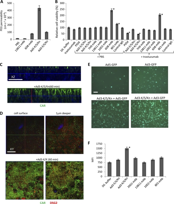
Fig. 7.
Functional analyses of epithelial junction opening. (A) T84 cells were grown in polyester membrane transwell inserts for 21 days until transepithelial resistance was constant, implying that tight intercellular junctions had formed. Shown is [14C]PEG-4000 diffusion through polarized T84 cells cultured in transwell chambers. Cells were incubated with PBS or the various DSG2 ligands for 15 min, after which [14C]PEG-4000 was added to the inner chamber. Paracellular flux was assessed in aliquots from the apical and basal chambers as described elsewhere (1). The following monoclonal antibodies against different extracellular domains (ECD) of DSG2 were used: 13B11-MAb (against ECD 1 and 2) and 6D8-MAb (against ECD 3 and 4). Ad3-K/S/Kn is the dimerizing form. Ad3-E/S/Kn is unable to dimerize. (B) Effect of DSG2 ligands on trastuzumab killing of Her2/neu-positive breast cancer BT474-M1 cells. Cells were incubated at 100% confluence for 2 days. Ligands were added to the inner chamber for 1 h, followed by PBS or trastuzumab. Cell viability was measured 2 h later (see Materials and Methods). In addition to 13B11 and 6D8 MAbs, the following anti-DSG2 antibodies were used: 7H9 (against the propeptide domain), 10D2 (against ECD 2), and 8E5 (against ECD 3 and 4). The viability of dilution buffer-treated cells was taken as 100%. n = 5, i.e., five separate wells. *, P < 0.05 compared to data for dilution buffer. (C) Confocal microscopy for CAR in T84 cells treated with dilution buffer or Ad3-K/S/Kn (stacked x-z layers). T84 cells were grown in polyester membrane transwell inserts for 21 days. Ad3-K/S/Kn (40 μg/ml) or dilution buffers were added for 60 min, after which cells were washed and subjected to immunofluorescence analysis. CAR appears as green staining. Nuclei are blue. The scale bar is 20 μm. (D) Confocal microscopy for CAR in polarized T84 cells. x-y images were taken from the cell surface and at a layer 1 μm beneath the cell surface (right). Experimental conditions were the same as those for panel C. The scale bar is 20 μm. (E) Ad transduction of T84 cells. T84 cells were grown in polyester membrane transwell inserts for 21 days. Ad3-GFP and Ad5-GFP were added to the inner chamber at an MOI of 250 PFU/cell together with dilution buffer (upper) or Ad3-K/S/Kn (40 μg/ml). Three hours later, virus was removed and cells were washed. GFP expression was analyzed after 20 h of incubation. Shown are representative images. For quantification, GFP-positive cells from 10 independent images of three independent experiments were counted. The scale bar is 20 μm. (F) Flow cytometry of Ad5-GFP-infected cells. T84 cells were infected with Ad5-GFP as described for panel E either in dilution buffer or 40 μg/ml DSG2 ligands. n = 3. *, P < 0.05 compared to data for dilution buffer.

