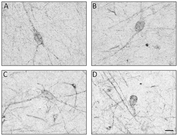
Figure 2.

High-power photomicrographs of 5-HT1A-immunoreactive cells in the hippocampus of Braak 0–II cases. Cells with morphological characteristics of pyramidal (A), bipolar (B), or multipolar (C) neurons and interneurons (D) exhibit punctate 5-HT1A immunoreactivity at the level of cell membranes of soma and dendrites. Scale bar = 20 μm.
