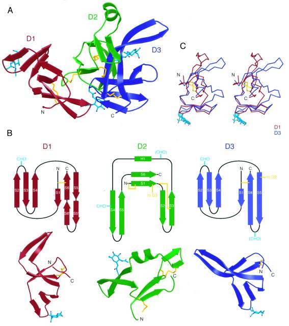
Figure 2.
Structure of the Mth ectodomain. (A) Ribbon diagram of the Mth structure (D1, red; D2, green; D3, blue). Ordered N-linked carbohydrates are shown in cyan in ball-and-stick representation and disulfide bonds are yellow. (B) (Upper) Topology diagrams for three extracellular domains of Mth. Potential N-linked glycosylation sites are labeled CHO, with parentheses denoting N-linked sites in which ordered carbohydrates were not observed. (Lower) Ribbon diagrams of the three domains of Mth. A disulfide bond (yellow) and an N-linked carbohydrate (cyan) are located at corresponding positions in D1 and D3. (C) Stereoview of a superposition of D1 (in red) and D3 (in blue) made by aligning the Cα atoms of the S2-S3-S4 β-sheet.

