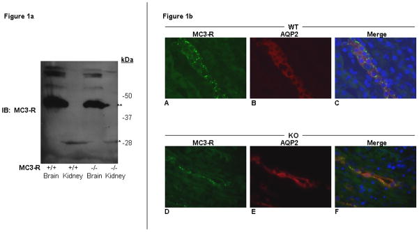
Figure 1. MC3-R protein is not specifically detectable in mouse brain and kidney.
a) Western blot analysis of brain (Brain) and kidney (Kidney) homogenates from wild type (+/+) and MC3-R knockout (−/−) mice. Blots were probed with a rabbit anti-MC3-R antibody (Sigma Aldrich M4937). A ~40Kda band (**) is seen both in the +/+ and −/− samples of mouse brain tissue. A smaller ~28 kDa band (*) is seen in the +/+ and −/− kidney lanes. b) Immunohistochemistry of MC3-R wild-type (WT) and knockout (KO) mouse kidney sections. Sample sections from WT (A–C) and KO (D–F) mouse kidney stained with a rabbit anti-MC3-R antibody (Sigma M4937) and a goat anti-AQP2 antibody (Santa Cruz Biotechnology C-17). Images stained for MC3-R (A and D), AQP2 (B and E), and merge with DAPI staining (C and F).

