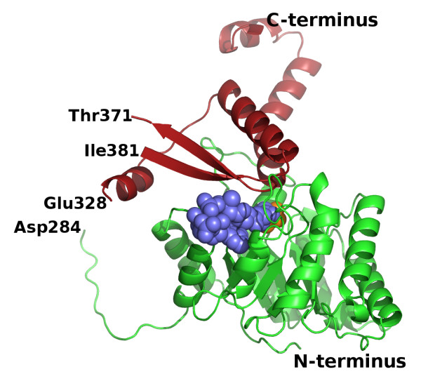
Figure 1.
Overall fold of an H. volcanii MSH monomer in the ternary complex. The N-terminal β8/α8 barrel and the C-terminal domain are shown as cartoon ribbon traces in green and red respectively. Acetyl-coenzyme A and pyruvate are shown as space-filling models in slate blue and orange respectively. The protein segments between Asp284 and Glu328, and between Thr371 and Ile381 were not visible in the crystal structure and have not been included in the model.

