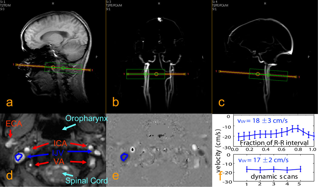
Fig. 2. Typical localizer images and blood velocity measurements for the internal jugular veins (IJVs).
a: anatomical scout image. b, c: angiographic scout images obtained using phase contrast MRA. The orange rectangle indicates the imaging slice for blood velocity measurements chosen perpendicular to the IJV. The green box is the localized shimming box (linear shims only). d: anatomical image of the selected slice, showing IJVs, internal carotid arteries (ICAs), external carotid arteries (ECAs), and vertebral arteries (Vas). e: quantitative velocity map (units of cm/s) for the selected slice. f: velocities measured with PPU triggering (top) and without triggering (bottom) for one of the healthy adult volunteers, showing good estimate of the average velocity with dynamic scans (bottom) even with some pulsatile flow (top) in IJV.

