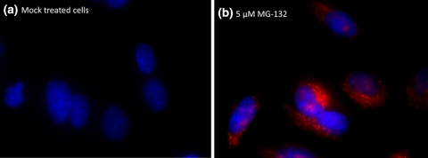
Fig. 2.
Protein aggregation detected by ProteoStat® dye as observed by fluorescence microscopy: Hela cells were mock-induced with 0.2% DMSO (a) or induced with 5 μM MG-132 (b) for 16 h at 37°C. After treatment, cells were incubated with ProteoStat® dye for 30 min. Nuclei were counter-stained with Hoechst 33342 in the images

