
Figure 1.
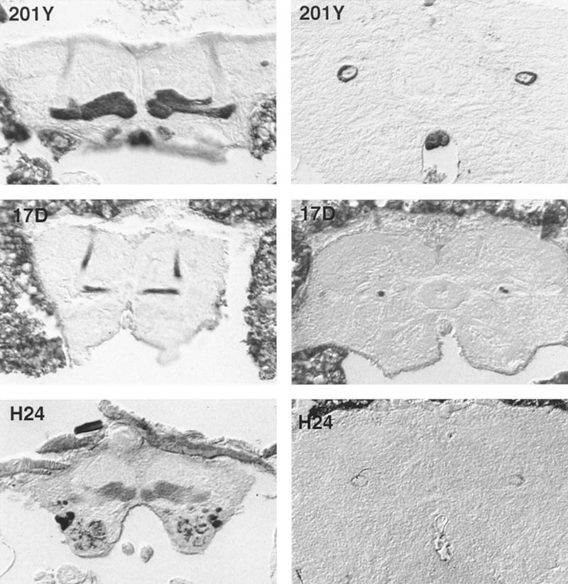
Tetanus toxin light chain staining in adult brains of the three groups of P[GAL4]/Cnt-E flies used in this study. The tetanus toxin expression is revealed by anti-tetanus toxin antibody. The expression pattern correlates well with that of other reporter genes (e.g., β-galactosidase; data not shown). Cryostat frontal sections (10 μm) at the level of the α- and β/γ-lobes (left) and peduncule cross-sections at the level of the CC (right). Line P[GAL4]201Y exhibits an extensive staining in the γ-lobe, whereas only faint staining is observed in the α- and β-lobes. In the peduncule, most of the staining is restricted to an outer ring (γ-fibers). The small dot in the center may correspond to the immunopositive fibers of the α- and β-lobes (α/β-fibers). In line P[GAL4]17D staining is found in the α- and β-lobes but not the γ-lobes. In the peduncule, staining is restricted to the central core, absence of staining in outer ring correlates with absence of γ-lobe staining. Line P[GAL4]H24 shows, in the MBs, a staining only in the γ-lobe. The faint staining restricted to the periphery of the peduncule corresponds to the γ-lobe staining. The difference in the size of the stained patterns between the three P[GAL4] lines is attributable to a difference of head sizes.