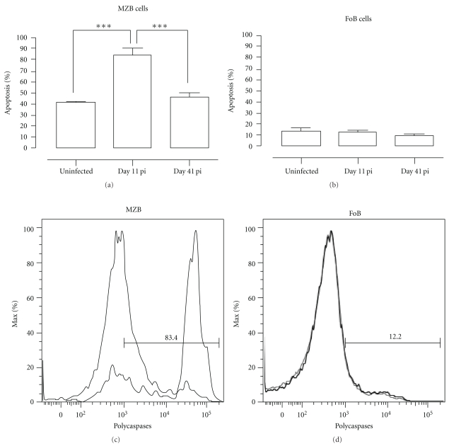
Figure 8.
PcAS infection-induced apoptosis of MZB but not FoB cells. Spleens cells from noninfected mice and mice infected with PcAS for 11 and 41 days were stained for surface markers commonly used to define MZB and FoB cells and the amount of active caspase 1, -3, -4, -5, -6, -7, -8, and -9 was measured intracellular by flowcytometry. (a) and (b) Percentage of apoptotic cells within MZB or FoB cell population in uninfected controls versus infected mice on day 11 and 41 pi. Data are represented as mean of three mice per group ± SEM (***) P < .001. (c) and (d) Representative histogram of uninfected controls (grey line) versus a day 11 pi (black line).

