
Figure 1. Different components of deep DHN receptive field.
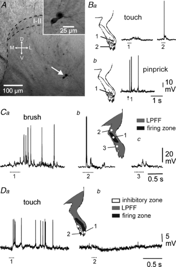
A, cell body location of biocytin-labelled, patch-clamp recorded deep DHNs. In this particular case, two neurones appeared dye-coupled raising the possibility of gap junction coupling (inset). I–II, laminae I–II; M, medial; L, lateral; D, dorsal; V, ventral. B, selected cells are wide dynamic range (WDR) neurones, responding to both innocuous (touch) and noxious (pinprick) stimulations (Ba and b, respectively). Part of the receptive field may be subthreshold for touch (Ba, 1), and suprathreshold for pinprick (Bb, 1). C, DHNs present a firing zone (inset, black area) in which innocuous stimulation leads to a burst of spikes (Ca, 1) surrounded by a low probability firing fringe (inset, grey area). D, most DHNs have both a suprathreshold excitatory receptive field (Da, 1) and inhibitory zones (Db, 2). At resting potential, individual IPSPs are barely visible. B and C, same deep DHN. D, superficial DHN.