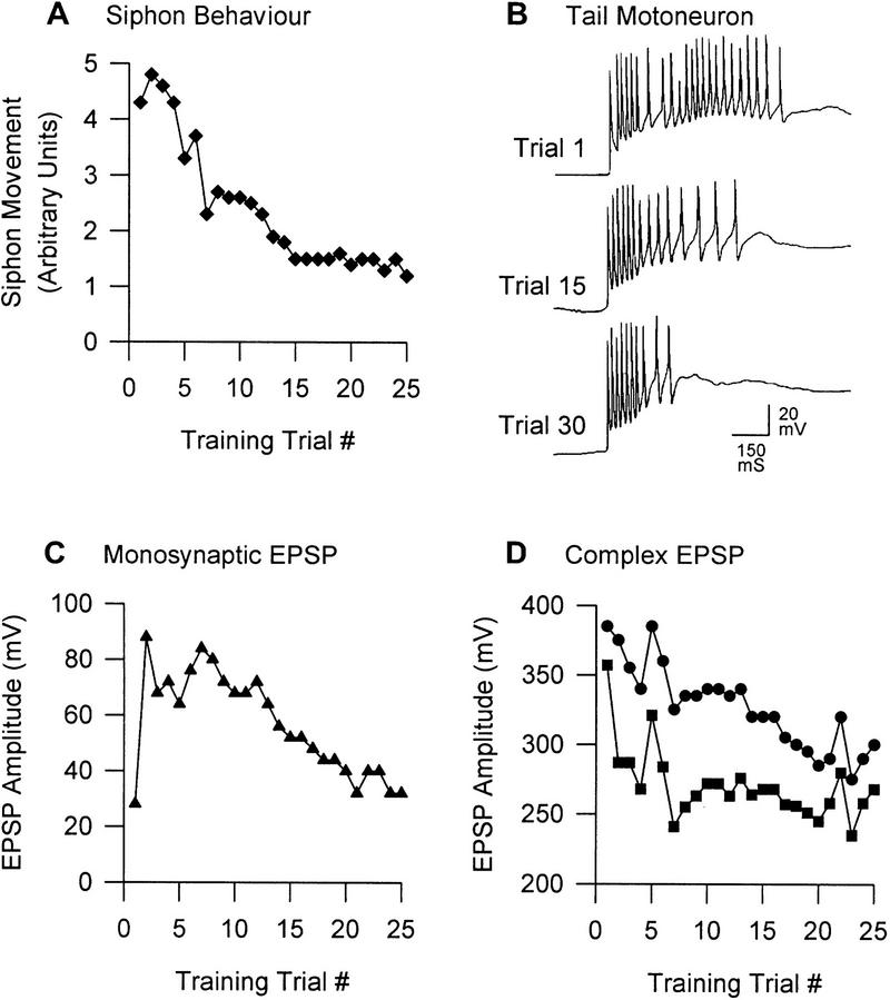
Figure 5.
Tail-elicited withdrawal reflexes in Aplysia. (A) Plasticity of the tail-elicited siphon withdrawal response. This behavior exhibits transient sensitization followed by habituation; these behavioral changes are parallelled by changes in the number of action potentials in siphon MNs (not shown here; see Stopfer and Carew 1996). Stopfer and Carew (1996) also show similar changes in tail-elicited tail MN activity and the tail withdrawal response, though the behavioral changes are quite erratic. (B) Tail MN activity. Short-latency spiking is mediated by monosynaptic pathways, whereas longer latency spiking is mediated by polysynaptic pathways (White et al. 1993). Note the dissociation of plasticity between these two pathways; between trials 1 and 30, the monosynaptic pathway exhibits facilitation, whereas the polysynaptic pathway exhibits depression. A strikingly similar dissociation of plasticity between the different phases of the neuronal response is seen in the S neurons of the spinal cat (Groves and Thompson 1970). (C) Monosynaptic EPSP in tail MN. When the tail MN is prevented from spiking, the monosynaptic EPSP shows sensitization consistent with the data in B but eventually habituates. (D) Complex EPSP in tail MN. The complex EPSP, consisting of monosynaptic and polysynaptic components (•), does not change analogously to the monosynaptic EPSP (C). If the monosynaptic component is subtracted (based on the assumption of linear additivity and four monosynaptic inputs; see White et al. 1993), one sees the polysynaptic component (█) that appears to exhibit fairly pure depression. Adapted, with permission, from Stopfer and Carew (1996).

