
Figure 1.
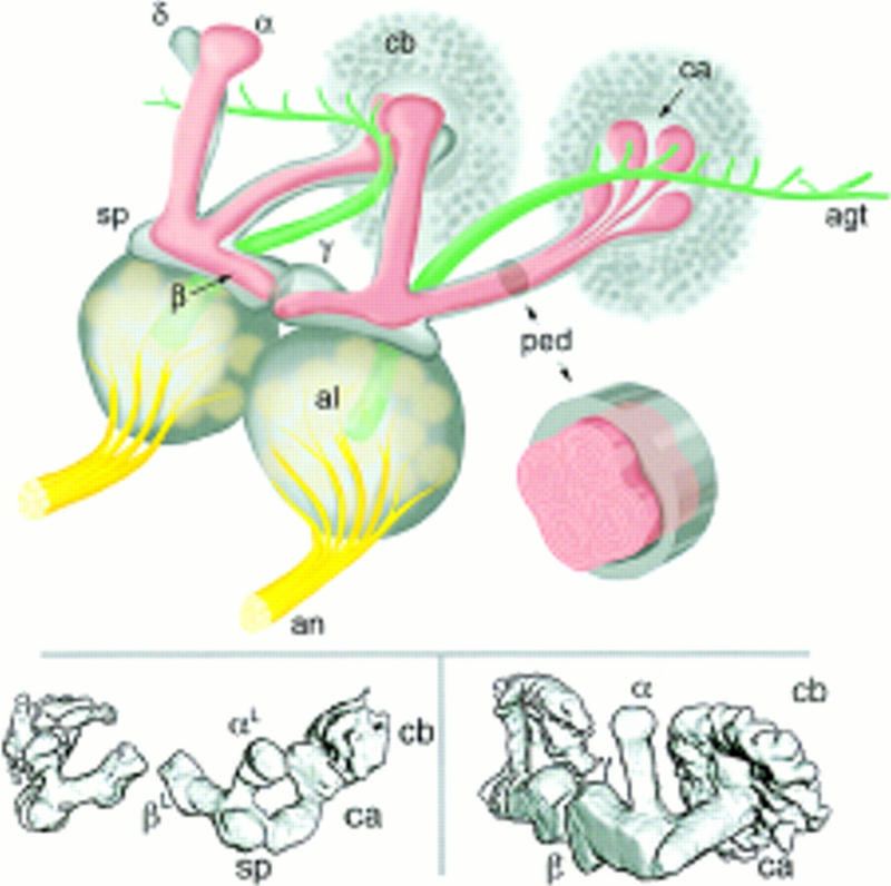
Drosophila mushroom bodies. (Top) Schematic representation of the adult Drosophila olfactory system. Olfactory input from sensory receptors on the antennae reaches the antennal lobes (al) via the antennal nerve (an). The glomerular arrangement of the AL is indicated. The AL also receives olfactory input from the maxillary palps (not shown). Among the outputs of the AL is the antennal glomerular tract (agt), projecting caudally toward the MB calyx (ca), and then to the lateral protocerebrum. The calyx contains the dendritic arbors of KCs and is half contained within a rind of KC cell bodies (cb). KC axons arising within the calyx project rostrally via a stalk-like structure known as the pedunculus (ped). Upon reaching the rostral margin of the central brain, the pedunculus gives rise to a spur-shaped lateral projection (sp), and to four lobes (α,β,γ,δ). β and γ project toward the midline, where they almost abut. α and δ project to the dorsal margin of the brain. MBs exhibit a clear and continuous fourfold symmetry within the cell body layer (four adjacent “clouds”), the calyx and the pedunculus (four internal tracts, each of which has a concentric organization). As they transit the pedunculus the four tracts merge, and it is their projections that form the α and β lobes. Surrounding the four peduncular tracts is a sheath of KC fibers projecting via the spur to form the γ lobe and at least some elements of the δ lobe. The inset is a schematic representation of a cross-section though the pedunculus, illustrating its internal organization. (Bottom) Surface-rendered models of late third instar larval (left) and adult (right) MBs as visualized in P{GAL4} line 201Y. The main MB structures are indicated, with the exception of the adult δ lobe, which is not encompassed by the expression pattern of line 201Y.