
Figure 2.
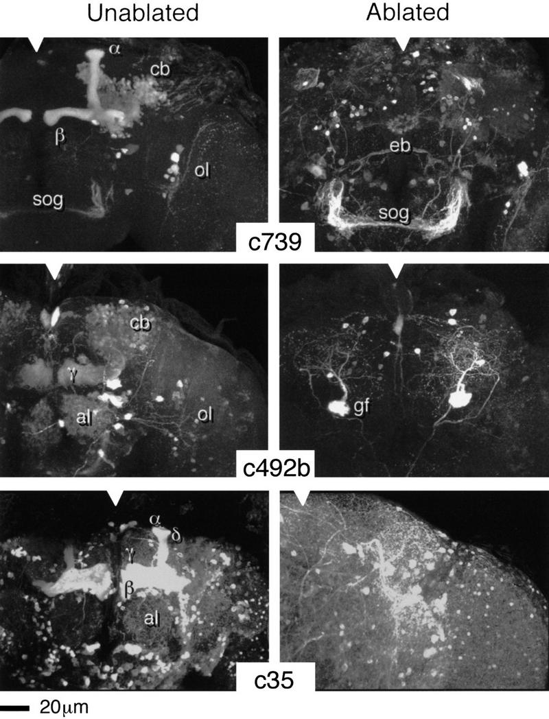
HU ablation of postembryonic KCs. Three-dimensional reconstructions of adult brains from the indicated P{GAL4} lines. (Notches) Approximate position of the midline in each panel. (c739) The normal expression pattern reveals the α and β lobes of the MBs, intrinsic neuronal components of the ALs (weak), the ellipsoid body—a component of the central complex (weak), together with elements of the optic lobes (ol) and the subesophageal ganglion (sog). (c492b) The normal pattern reveals large numbers of KCs within the α, β, and γ lobes (α and β less clear in this reconstruction), intrinsic neurons of the ALs (al), the giant fiber (gf, partly obscured by MB staining), and elements of the optic lobes. (c35) The normal pattern extends widely through central brain neuropil but is strongest within the MBs (α, β, γ, and δ). In each case, the ablated pattern is selectively and completely depleted for MB and AL staining. Surrounding neuropil, as far as can be determined, appears normal.