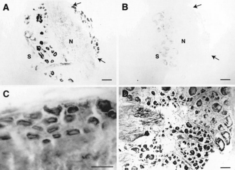
Figure 2.
TβR-II immunoreactivity in sections of pleural and pedal ganglia. (A) Staining is present in neuronal cell bodies in the pleural ganglia as well as in the neuropil, which may represent staining along neuronal processes. The cluster of mechanoafferent sensory neurons in the ventral–caudal cluster also exhibit immunoreactivity and can be identified by size and position (area between arrows). (S) Sheath; (N) neuropil. (B) Control section adjacent to that in A shows little staining. (C) Higher magnification view of mechanoafferent sensory neurons from a different section than that shown in A. (D) Immunoreactivity is also present in many cells of the pedal ganglion, particularly in the caudal region, shown in this section. Scale bars, 100 μm (A,B,D) and 50 μm (inset).

