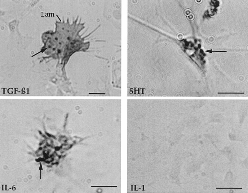
Figure 2.
Immunocytochemistry of hemocytes migrating on polylysine-coated plastic dishes. The cells were fixed, permeabilized, and exposed to the primary antibody indicated. Antigen-antibody complexes were subsequently detected with HRP-coupled secondary antibody. Hemocytes were stained by antibodies to vertebrate TGFβ1 and IL-6, but not to IL-1. Positive staining was also obtained with an antibody to 5-HT. The staining was most intense in large granules (arrows). Hemocytes that were recognized by the anti-TGFβ1 antibody were larger than most of the other cells and contained especially prominent lamellipodia (Lam). Bars, 2 μm.

