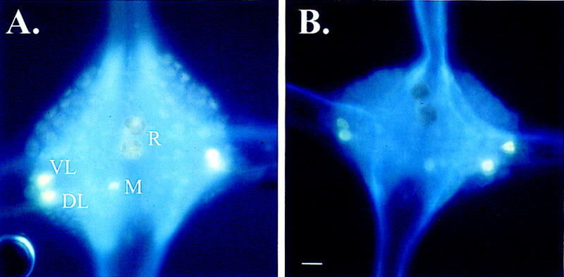
Figure 1.

Glyoxylic acid staining of 5-HT-depleted and control ganglia. (A) Ganglion from a leech treated with ascorbic acid. In these control animals, the R cells do stain positive for 5-HT. Other serotonergic neurons that are stained by this procedure are the DL, VL, and posteromedial (M) neurons. (B) Ganglion from a leech treated with 5,7-DHT. Although other 5-HT-containing neurons still stain positive for 5-HT, the R cells do not stain for 5-HT indicating depletion of 5-HT. Bar, 50 μm.
