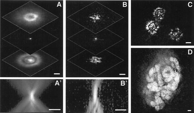
Figure 1.
The problem: Sample-induced optical aberration due to heterogeneous refractive index. (A) An example of a nonaberrated 3D image of 0.1-μm bead using high magnification (100×/NA1.3 objective) microscopy. In-focus and +/− 3 μm defocus images are shown. The subpanel A' is an xz section through the center of the 3D bead image. (B) A fluorescent bead imaged three-dimensionally (as in A) under a polytene nucleus in a Drosophila salivary gland cell, showing sample-induced distortions. B' is the corresponding xz section. (C) The image of a field of view containing several beads under a polytene nucleus, showing the spatial dependence of these distortions. The image is taken slightly out of focus to emphasize the different distortions. (D) DNA-specific dye Oli Green staining of polytene chromatin in a live cell. The optical section shown was processed by deconvolution using the symmetric unaberrated PSF. The familiar chromosome bands are almost entirely obscured by distortions for both the unprocessed or processed image. (Scale bars, 2 μm.)

