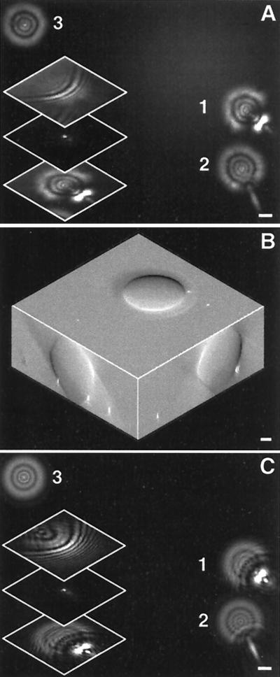
Figure 3.
Proof of principle: (A) Submicron beads on a slide were imaged under an oil droplet (18 μm in diameter). Distorted aberrated images were recorded. In the Inset, three optical sections for the bead 1 display a strong out-of-focus “flare” and highly asymmetric out-of-focus diffraction pattern (arcs instead of Airy rings). (B) The DIC images of this sample recorded three-dimensionally. Orthogonal section views are presented, with the superimposed projected positions of the beads. (C) PSFs computed by ray tracing through the oil droplet for the positions of the beads. The insert is three optical sections calculated for the bead 1. (Scale bars, 2 μm.)

