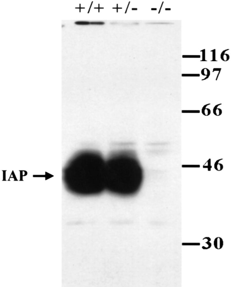
Figure 3.
Representative gel pattern showing the results of Western blotting of IAP expression in the whole brain of IAP+/+, IAP+/−, and IAP−/− mice. IAP monoclonal antibody (miap301; Lindberg et al. 1996b) was used. IAP protein was absent in IAP−/− mice and IAP expression was similar in both IAP+/+ and IAP+/− mice.

