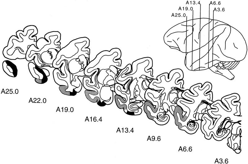
Figure 2.
Line drawings of representative coronal sections through the temporal lobe of M. fascicularis adapted from the atlas of Szabo and Cowan (1984). The sections are arranged from rostral (A25.0) to caudal (A3.6), and the rostrocaudal position of each section is indicated in the lateral view. The designations A25.0, A22.0, and so on, specify distances anterior (A) to the intra-aural line. The boundaries of the perirhinal cortex are indicated in black, and the boundaries of area TE are indicated in gray.

