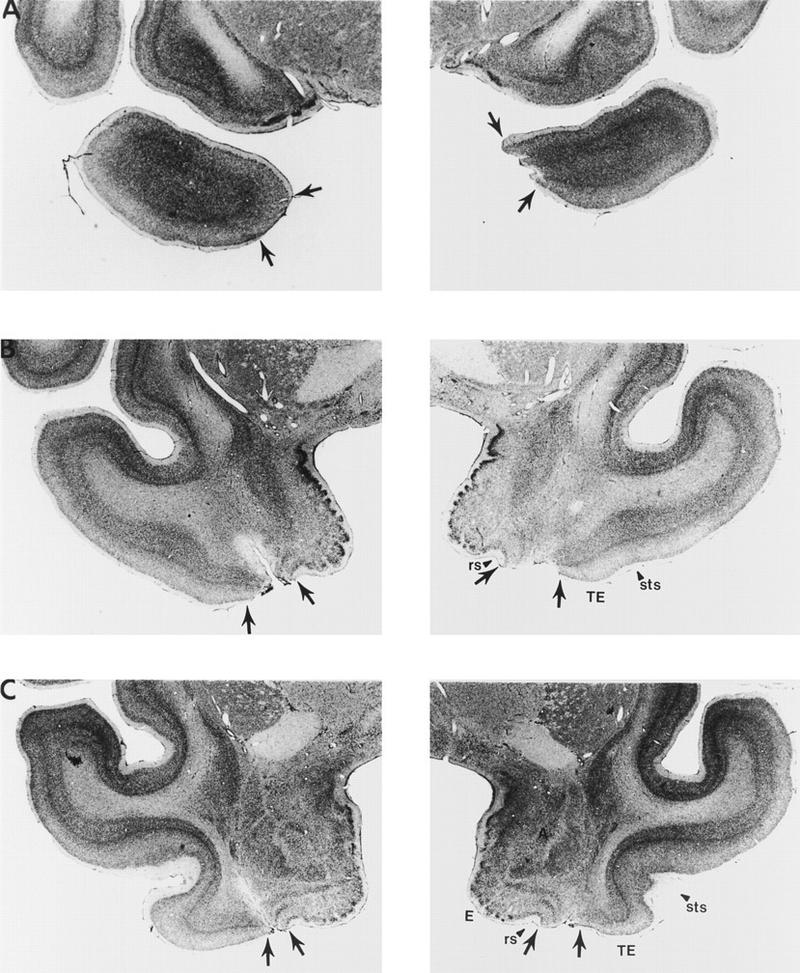
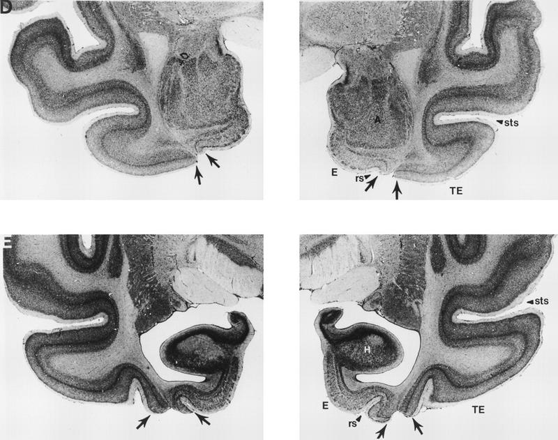
Figure 4.


Photomicrographs of representative sections through the left and right temporal lobes of monkey PR 2, whose lesion most closely approximated the intended lesion (see Fig. 2). The sections are arranged from rostral (A) to caudal (E), and the lesion is indicated by arrows at each level. (rs) Rhinal sulcus; (sts) superior temporal sulcus; (TE) area TE; (A) amygdala; (E) entorhinal cortex; (H) hippocampus.
