
Figure 3.
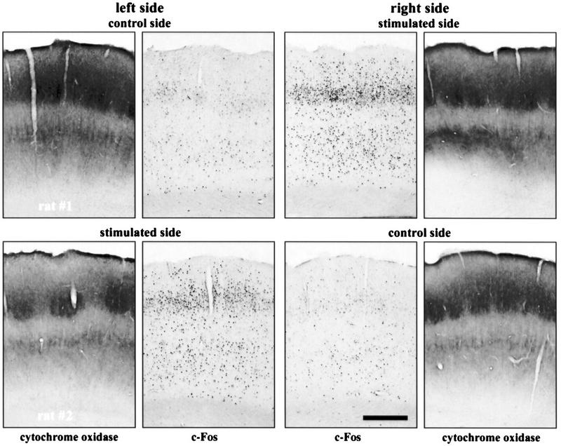
Immunocytochemistry showing an increase in c-Fos expression in the PMBSF corresponding to stimulated vibrissae (experiment 1). Shown are both left and right PMBSF of two rats that had their whiskers brushed on the left side (top) and the right side (bottom) of the snout. Pictures of cytochrome oxidase staining of neighboring sections are included. Calibration bar, 0.5 mm.