
Figure 3.
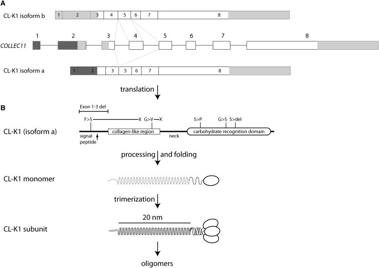
Gene Structure, Alternative Splicing, and Protein Products of COLEC11
(A) Overview of the gene/primary transcript and the two suggested alternative splice variants. Mutually exclusive splice events are indicated by the dotted lines. Greyed areas indicate the 5′ and 3′ UTRs. Notice the predicted use of alternative translation initiation sites in isoforms a and b.
(B) The protein structure of the secreted isoform is illustrated and polymorphisms are indicated, along with the mutations reported by Rooryck et al. The exact locations of the mutations are: c.496T>C (p.Ser169Pro), c.610G>A (p.Gly204Ser), c.45delC (p.Phe16SerfsX85), c.648_650delCTC (p.Ser217del), and c.300delT (p.Gly101ValfsX113), as well as the 27 kb exon 1–3 deletion. The formation of the structural subunit composed by three identical polypeptide chains and the ensuing oligomerization is illustrated.