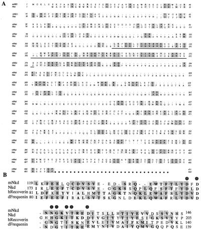
Figure 1.
Sequence comparison of mNkd and the Drosophila Nkd. (A) Protein sequence alignment of mNkd and Drosophila Nkd. Deduced protein sequences of mNkd and Nkd (17) are compared with the Macvector clusterw program. Identical residues are highlighted in dark gray; conserved changes are highlighted in light gray. The EF-hand motifs and the surrounding amino acids in both proteins are underlined. (B) Alignment of the underlined amino acids of mNkd, Nkd (17), human Recoverin (41), and Drosophila Frequenin (42). Residues that are identical in more than two proteins are highlighted in dark gray; conserved changes are highlighted in light gray. Consensus residues in the EF-hand that bind calcium with high affinity are indicated by shaded dots above these residues (17, 20).

